


Chloroplast Density
For this part of the lab we're going to take a closer look at chloroplasts, and relate that information to cell structure, function, and evolution. Our model organism for this activity is the lace plant (Aponogeton madagascariensis), so named for its unique leaf morphology. The characteristic holes in the leaves of the lace plant are due to a process called programmed cell death, or apoptosis, where cells die "on purpose" during the development of the organism or structure. The lace plant is an emerging model species for cell death research, due to this highly predictable cell death pattern, and to its thin transparent leaves which allow for extensive microscopic observation of cellular activity throughout development. There are a few hypotheses about why the lace plant forms these holes, such as an anti-predation/camouflage strategy, or to decrease water resistance. The ultimate reason, however, remains a mystery (perhaps one you will solve some day?). Although it is an endangered species in the wild, its natural beauty and uniqueness has made it a popular cultivated plant not only for scientists, but for aquarium hobbyists as well.
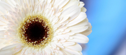
Lace plant (Aponogeton madagascariensis)

botanical illustration by Sir William Jackson Hooker (1856)

A. madagascariensis in an aquarium.
Data Collection
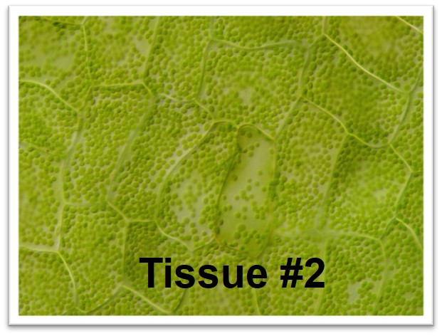
The images below are micrographs of cells from two different parts of the lace plant. Click on the photos to open up larger versions, save a copy of each large version to your computer.
Use Fiji/ImageJ (see the Downloading, Installing, and Testing Fiji/ImageJ page in this lab) to count the chloroplasts in five different cells from each photo (identified for you on the larger versions). Enter your data into the table in your lab document and answer the associated questions.
.jpg)
The last question for this section in your lab document refers to the labelled photo of a lace plant below. (The sheath is a protective layer of cells found around the base of the stem, and the corm is an underground shoot.)
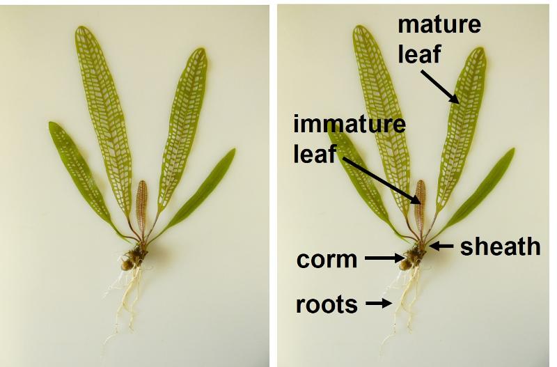
Image Credits
'Tissue 1', 'Tissue 2', and lace plant whole mount photos by Jacob Fletcher
lace plant illustration by Sir William Jackson Hooker (1785 - 1865) [Public domain], via Wikimedia Commons
lace plant aquarium photograph [CC0] via Pixabay